St. Mary's High School, Manhasset, NY
|
** Important Disclaimer ** |
|||||||||||||||
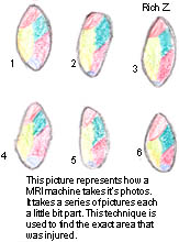
MRI
![]() INTRODUCTION - Written by, Nicole L.
INTRODUCTION - Written by, Nicole L.
A MRI (magnetic resonance imaging) is a very useful and helpful technique. It is useful in detecting small tumors, blocked blood vessels or damaged vertebral disks. Because it does not use radiation, it can be used where x-rays are too dangerous. The way an MRI works is pretty simple. Large magnets beam energy through the body causing hydrogen atoms in the body to vibrate. This produces energy in the form of tiny electrical signals. A computer detects these signals, which vary in different parts of the body and according to whether an organ is healthy or not. The variation enables a picture to be produced on a screen and is interpreted by a medical specialist. The concept and idea of using an MRI to detect tumors was proposed by Raymond Damadian in 1927. But the fundamental of MRI imaging concept used in all present day MRI instruments was proposed by Paul Lauterbar in 1973.

A MRI is very important to many people. Just by going for a simple testing by a MRI can save your life. A MRI can detect life-threatening diseases, tumors, etc. Basically, MRI "sees" the difference between life and death in great detail. The doctor uses a MRI Scan to diagnose brain tumors, abscesses, swelling, bleeding, nerve damage and other disorders that increase the fluid content of tissues. The test also shows irregularities of the spinal cord. Doctors use this advice when they are either uncertain of the problem or if and x-ray would be too dangerous towards the patients problem. The test only takes about an hour and a half to complete. Although, MRI is painless and involves no exposure to radiation from the scanner, a radioactive contrast dye may be used. The dye is injected near the organ or tissue being observed. MRI scanners are very small and deep. These MRI scanners are found in particular doctors' offices and in some hospitals.
![]() PART TWO - Written by, Mayrose C.
PART TWO - Written by, Mayrose C.
Magnetic Resonance Imaging is also known as MRI. It is a medical tool used for diagnosis. It is based on the principles of Nuclear Magnetic Resonance imaging. A patient is placed inside a cylinder that contains a strong magnet. Radio waves are then placed into the cylinder, which causes hydrogen in the body to vibrate. It does not use ionizing radiation. It can see through bones and produces images of blood vessels, cerebrospinal fluid or the liquid in the spine and brain, cartilage, bone marrow, muscles and ligaments. It is a harmless procedure except for persons with metal objects implanted in their bodies, such as pacemakers, joint pins, or artificial heart valves. The powerful magnetic fluid may dislodge these objects. A radio frequency is sent in and replaces the vertically aligned proton by 90 degrees in the horizontal plane. When the RF (radio frequency) pulse is turned off the proton will go back to their original position. The faster they return to the original position, the stronger the signal and brighter the visualized structure. When the RF pulse was turned off the protons started to change direction at different rate.
The main magnet is located in the large donut -shaped structure in which the patient lies. The main magnetic field is kept constant. Its strength is measured in units called Tesla or T and ranges from 0.08 T to 2 T.
The gradient system is usually compromised of three electromagnets that can be turned off easily. It is used to produce a small magnetic field that can select, slice and encode positions in the image, when added to the main field. In order to form an image, the source and strength of a signal must be determined. Gradient field is a weak magnetic field, which is lain over the main magnetic field.
The MRI RF transmitter is similar to that used in radio stations. RF coils or the antennae that transmit and receive radio waves. There are two types: The larger volume coils which surround the body part being imaged and the surface coils which are placed directly under the area of interest and provide an intense detail.
BIBLIOGRAGHY
" MRI Indications for the referring physician. " Basic Image Interpretation, MRI Hardware. Online, Internet. March 1998.
"Rayden" . Macmillan Encyclopedia of Physics. 1996 ed. Volume III .
Newhouse, J.H. Understanding MRI. (1991) Oldendorf, W.H. Mri Primer (1991).